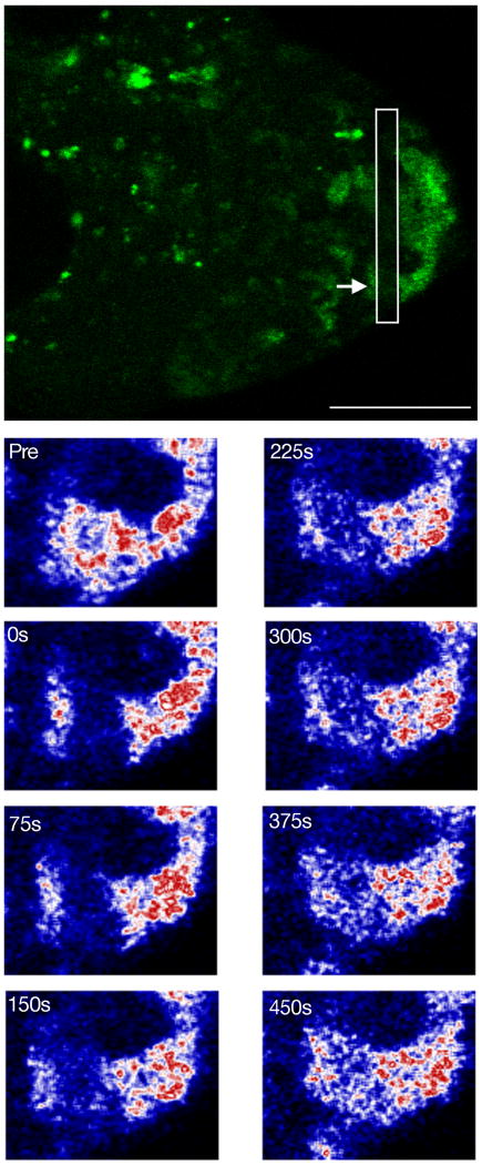
Fig. 6. Exchange of Stau between reticulated bodies and cytoplasm.
The top panel shows Stau∷GFP in a stage 8 egg chamber immediately after photobleaching. The white box surrounds the photobleached region and the arrow indicates the photobleached reticulated body shown in the bottom panels. The photobleached region is close to the posterior of the oocyte. Scale bar is 20 μm.
The lower panels are of Stau protein in a close up of a reticulated body before bleach (Pre) and at various times after photobleaching (times indicated in seconds). The oocyte posterior is to the right of the image. Stau∷GFP fluorescence is reacquired evenly throughout the photobleached region. The image of Stau∷GFP was artificially colored by converting its Look Up Table (LUT) from green to the Union Jack LUT in ImageJ (NIH). In this LUT color changes from black to blue to white to red with progressively higher fluorescence levels.

