Figure 1.
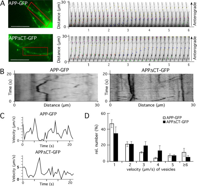
APP can be transported by the fast axonal transport machinery in the absence of its intracellular C terminus. Mouse primary cortical neurons (DIV7) expressing APP–GFP and/or APPΔCT–GFP were analyzed by time-lapse microscopy 18 h after transfection. A, Fluorescence micrographs of APP–GFP- and APPΔCT-GFP-expressing neurons. For tracking and velocity analyses, a region of interest (red box) was selected. Sequential images of the region of interest are shown to the right. Colored arrowheads indicate the single vesicles that have been examined. Time interval between images was 200 ms, 5 images/s. B, Representative kymographs showing APP–GFP and APPΔCT-GFP movement. C, Velocities of APP–GFP- and APPΔCT-GFP-containing vesicles, assayed over a period of 22 s. D, Histogram showing quantification of the number of recorded vesicles moving at velocities of 1, 2, 3, 4, 5, or >6 μm/s. No statistical significant differences between the velocities of APP–GFP- and APPΔCT-containing transport vesicles could be determined (Student's t test, p ≥ 0.2). Scale bar, 25 μm.

