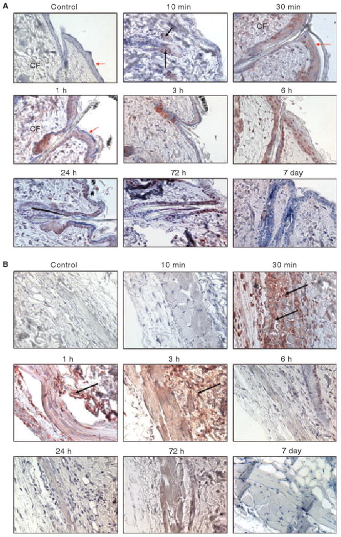
Fig. 1.

Immunostaining of skin tissues for rFVIIa in mice at different time intervals following the administration of a single pharmacological dose of rFVIIa (AF488-FVIIa). The sections were stained with anti-AF488 antibodies. Panel A, epidermis and upper dermis region; Panel B, lower dermis region. CF, collagen fibers. Red arrows point out squamous epithelial cells whereas black arrows point out hair follicles. Magnification, 400×. In this and other figures, control IgG staining was carried out with tissue sections of mice given AF488-FVIIa (30-min time period) or saline, where appropriate.
