
Fig. 3.
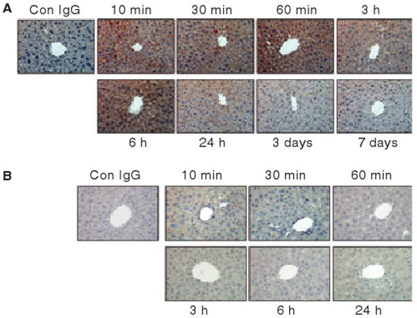
Immunostaining of liver sections with anti-AF488 antibodies of mice administered with AF488-FVIIa (panel A) or AF488-FIX (panel B). In panel A, the antibodies stained the portal vein, sinusoidal capillaries and hepatocytes. In panel B, only Kupffer cells were stained positively for AF488. Magnification, 400×.