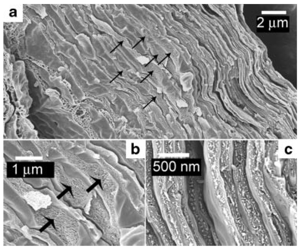
Figure 3.
Low and high magnification cryo-SEM images of the stratum corneum of natively hydrated porcine skin tissue. a: The stratum corneum consists of a dense outer layer and a less dense inner layer (each arrow marks the location of a corneocyte cell in the inner layer). b: The inner layers consist of irregular shaped corneocyte cells (indicated by arrows). Small gaps between corneocytes in b are artifacts of cryo-fracturing. c: The outer layer consists of densely packed flat sheets of corneocyte cells.

