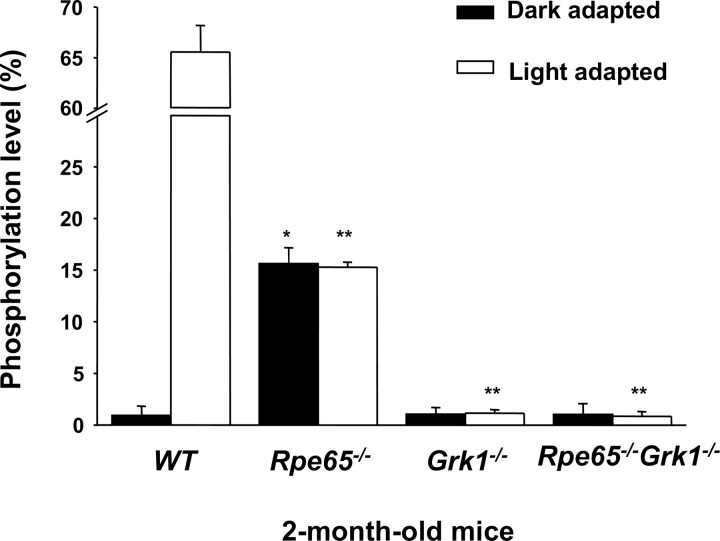
Figure 1.
Effect of deletion of Rpe65 and Grk1 on opsin phosphorylation. Retinae of cyclic-light-reared, 2-month-old WT, Rpe65−/−, Grk1−/−, and Rpe65−/−Grk1−/− mice were homogenized in 8 m urea and digested with endoproteinase Asp-N in 10 mm Tris buffer at pH 7.6 to cleave the opsin C terminus, which was analyzed online with an LCQ mass spectrometer. In the Grk1−/− and Rpe65−/−Grk1−/− mice, no significant opsin phosphorylation was observed. White bars, Animals exposed to room light for 6 h; black bars, animals dark-adapted for 12 h. Data are shown as the percentage of rhodopsin C terminus containing phosphorylation, independent of the multiplicity of phosphorylation, and presented as mean ± SEM; n = 3.

