
FIGURE 4.
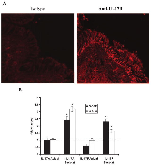
Detection and localization of the IL-17R. A, Representative immunohistochemical staining for IL-17R in a human lung section with a specific detecting monoclonal anti-IL-17R Ab, showing basolateral localization of the IL-17R. B, G-CSF and GRO-α secretion by primary HBE cells after addition of IL-17A and IL-17F (both at 10 ng/ml) to basolateral or apical surface. Basolateral media were collected after 24 h, and cytokine levels were measured by ELISA. Results are expressed as the mean ± SEM of triplicate samples from one representative experiment (*, p < 0.05).