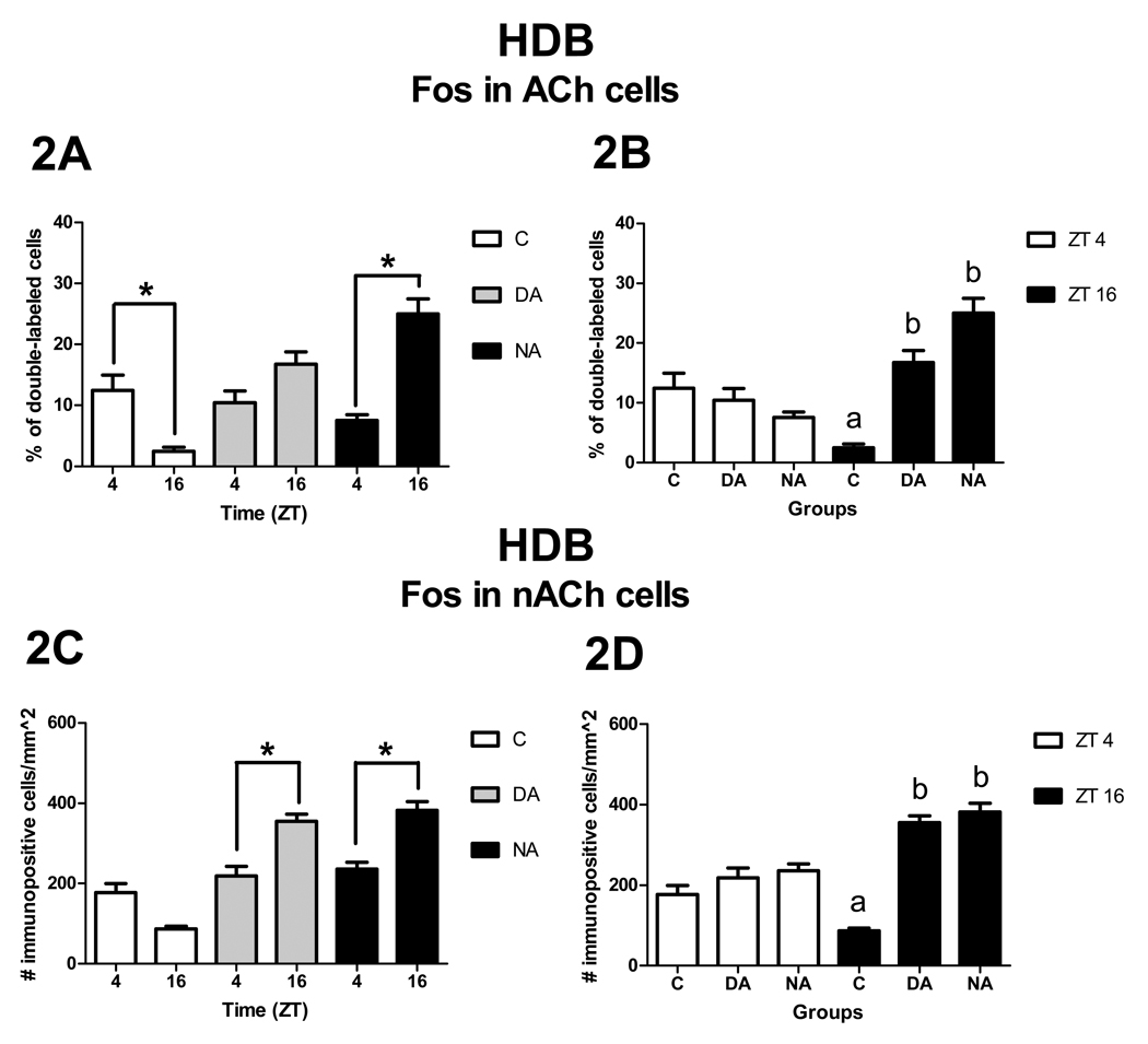
Figure 2.
Patterns of Fos expression in ACh cells (A, B) and nACh cells (C, D) in the HDB. Panels A and C show significant ZT differences (asterisks) within groups, whereas panels B and D show significant group differences within ZT (p< 0.05). Note that group means with different letters are significantly different from each other, here as well as in Figure 3, Figure 4, and Figure 7.

