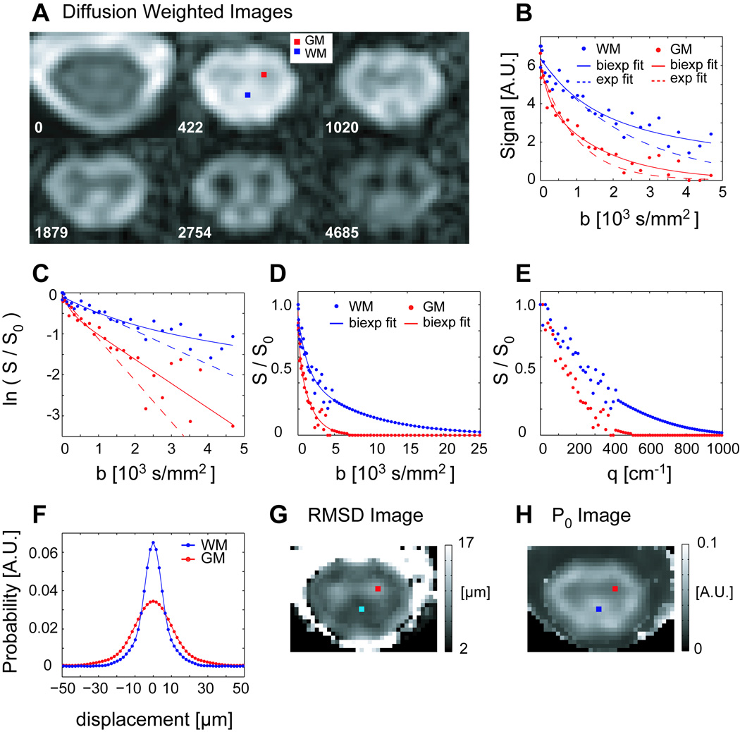
Figure 1.
A) DWIs (axial cross section, average of 2 diffusion weighting directions) through C4 spinal cord in a healthy 25 year old female volunteer. All DWIs (b>0) have the same windowing level. B) Measured signal decays for representative WM and GM voxels as a function of b-value, with bi-exponential (solid line) and mono-exponential (dashed line) fits. C) Natural log of the normalized signal showing departure from mono-exponential signal attenuation. D, E) The normalized signal, extrapolated and zero-padded (partial) data series as a function of b- and q- value, respectively. F) PDFs computed for GM and WM voxels. G, H) Maps of the RMSD and height (P0) of the PDF computed for each voxel at the slice level, with the location of the representative voxels indicated.

