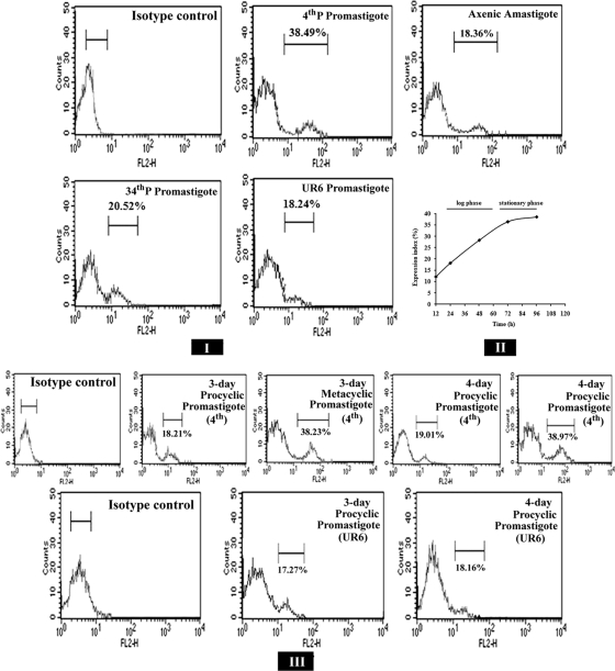
FIG. 2.
Flow cytometric analysis of expression of the 115-kDa extracellular serine protease. (I) Fluorescence histograms showing the expression levels of the 115-kDa protease in 4th-P, 34th-P, and UR6 promastigotes and axenic amastigotes of L. donovani. (II) Expression of the 115-kDa protease of 4th-P promastigotes of L. donovani at different phases of growth. The expression index measures the percentage of cells of the total population recognized by the anti-115-kDa antibody. (III) Fluorescence histograms showing the expression levels of the 115-kDa protease at procyclic and metacyclic stages of virulent promastigotes and at procyclic stages of attenuated UR6 promastigotes at 72 h and 96 h of culture.

