FIG. 1.
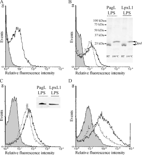
Incorporation of OpaJ protein and LPS derivatives into liposomes analyzed by flow cytometry and electrophoretic techniques. (A) Reactivity of anti-OpaJ MAb 15-1-P5.5 against OpaJ-containing liposomes measured by FCM analysis. (B) Reactivity of the anti-OpaJ MAb with liposomes containing LpxL1 LPS and OpaJ (continuous line) and with liposomes containing PagL LPS and OpaJ (dotted line). The inset shows the heat modifiability of OpaJ coincorporated with PagL or LpxL1 LPS into liposomes determined in seminative SDS-PAGE as a measure for its correct folding. Before electrophoresis, the samples were treated for 10 min in sample buffer with 0.1% SDS at room temperature (RT) or with 2% SDS at 100°C. The positions of molecular size markers are indicated at the left side of the membrane. (C) Reactivity of anti-L8 LPS MAb 43F8.10 with liposomes containing LpxL1 LPS and OpaJ (continuous line) and with liposomes containing PagL LPS and OpaJ (dotted line). The inset demonstrates the presence of LPS in the liposomes in an LPS-specific silver-stained Tricine-glycine-SDS-polyacrylamide gel. (D) Reactivity of the anti-L8 LPS MAb against liposomes containing PagL LPS and OpaJ in the presence (dotted line) or absence (continuous line) of the anti-OpaJ MAb. In all graphics, the reaction of antibodies with empty liposomes was included as negative control (gray-filled profile).

