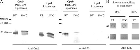
FIG. 2.
Binding of LpxL1 and PagL LPS to OpaJ protein. (A) Western blot patterns of liposomes containing OpaJ and PagL LPS or only OpaJ incubated with the anti-OpaJ MAb 15-1-P5.5 (left panel) or the anti-L8 LPS MAb 43F8.10 (right panel). Prior to SDS-PAGE, the samples were treated in sample buffer with 0.1% SDS at room temperature (RT) or with 2% SDS at 100°C. A complex of LPS and OpaJ is indicated in the right panel with an arrow. The positions of molecular size markers are indicated at the left side of the membranes. (B) Far-dot blots containing 15 μg of membrane-immobilized OpaJ that was denatured by heating for 30 min (100°C) or not (RT) prior to application on the membrane. The membranes were subsequently incubated with 500 μg of purified PagL LPS (a) or LpxL1 LPS (b) and then with the anti-L8 LPS-specific MAb 43F8.10. Bovine serum albumin (BSA) was used as a control for background binding.

