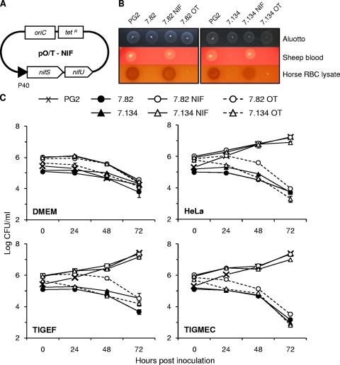
FIG. 5.
Complementation of M. agalactiae NIF mutants. Schematic representation of the plasmid pO/T-NIF used for complementation studies (A). The NIF locus was cloned under the control of the P40 protein promoter region. M. agalactiae parental strain PG2, mutants T07.082 (7.82) and T07.134 (7.134), and mutants transformed with plasmid pO/T-NIF (7.82 NIF and 7.134 NIF) or the control plasmid p21-1miniO/T (7.82 OT and 7.134 OT) were assessed for colony development on Aluotto, 10% horse red blood cell (RBC) lysates, or 5% sheep blood agar plates (B) and for survival over a 72-h incubation in DMEM-based medium (DMEM) or in coculture with HeLa, goat fibroblast (TIGEF), or goat epithelial (TIGMEC) cells (C). The data are the means of at least three independent assays. Standard deviations are indicated by error bars. Serial dilutions of mycoplasma stocks were spotted on blood agar plates, and colony development was observed after 4 to 6 days of incubation at 37°C.

