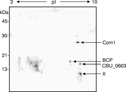
FIG. 1.
Representative 2D SW blot prepared from a total cell lysate of purified Coxiella SCVs (70 μg of total protein). IEF (pI, 3 to 10) was employed in the first dimension and a 12.5% (wt/vol) acrylamide SDS-PAGE gel was used in the second dimension, prior to blotting to nitrocellulose. The SW blot was probed with biotin-labeled, C. burnetii genomic DNA and visualized by streptavidin-horseradish peroxidase (see text for details). Potential DNA-binding proteins from the SW blot were identified by MALDI-TOF peptide mass fingerprinting and MALDI-TOF/TOF peptide sequencing and are listed on the right. The spot marked “X” could not be fingerprinted, despite numerous attempts.

