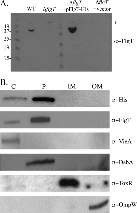
FIG. 6.
Cellular localization of FlgT. (A) Anti-FlgT immunoblot of periplasmic fractions isolated from V. cholerae WT, ΔflgT, ΔflgT/pFlgT-His6, and ΔflgT/vector strains. The asterisk indicates a cross-reactive band. The apparent molecular weights of the protein ladder are listed to the left of the gel. (B) Subcellular fractionation was carried out on the ΔflgT/pFlgT-His6 strain. The cytoplasm (lane C), periplasm (lane P), inner membrane (lane IM), and outer membrane (lane OM) fractions were isolated and separated by SDS-PAGE, followed by anti-His and anti-FlgT immunoblotting. Anti-VieA, anti-DsbA, anti-ToxR, and anti-OmpW immunoblots were performed to confirm fraction purity.

