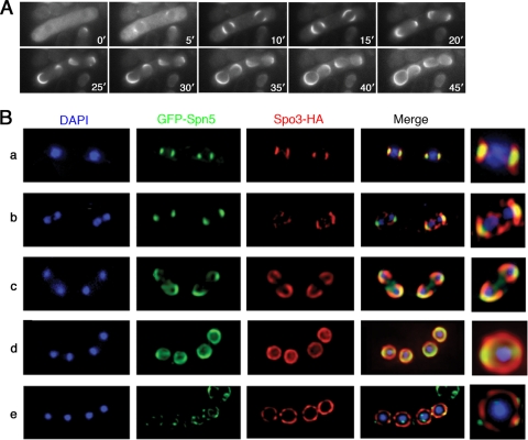
FIG. 8.
Formation of septin ring during sporulation via a horseshoe-shaped structure that extends along but is not coterminous with the FSM. (A) Wild-type strain THP18 harboring plasmid pREP41(GFP-spn2) was sporulated and observed by time-lapse microscopy. Times are indicated in minutes after the beginning of observation. (B) Wild-type strain THP18 harboring plasmids pREP41(GFP-spn5) and pAU(spo3-HA) was sporulated and observed by immunofluorescence with an anti-HA antibody (see Materials and Methods); Spo3 provides a marker for the entire FSM (53). Two-dimensional projections of three-dimensional deconvoluted images captured as 0.2-μm z sections were ordered according to the stages of meiosis II and spore formation, as follows: a, metaphase; b and c, anaphase; d, telophase; e, a mature ascus in which the Spo3-HA signal has begun to disappear. Magnified images of individual nuclear regions are shown on the right.

