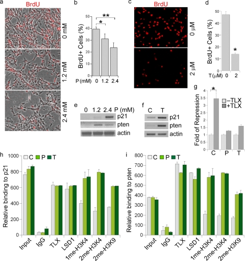
FIG. 2.
LSD1 inhibitor treatment inhibits neural stem cell proliferation. (a) Cell proliferation was revealed by BrdU labeling (red) in neural stem cells treated with different concentrations (0, 1.2, and 2.4 mM) of pargyline. The images shown are BrdU staining merged with phase contrast images. (b) Percentage of BrdU-positive (BrdU+) cells in 0, 1.2 mM, and 2.4 mM pargyline-treated neural stem cells. Error bars are standard deviations of the means; assays were repeated three times. *, P < 0.01; **, P < 0.001 (by Student's t test). (c) Cell proliferation was revealed by BrdU labeling (red) in solvent (0 μM)- and tranylcypromine (2 μM)-treated neural stem cells. (d) Percentage of BrdU-positive cells in solvent- and tranylcyptomine-treated neural stem cells. Error bars are standard deviations of the means; assays were repeated three times. *, P < 0.001 by Student's t test. (e) Gene expression regulated by the LSD1 inhibitor pargyline, revealed by RT-PCR analysis. Actin was included as a loading control. (f) Gene expression regulated by the LSD1 inhibitor tranylcypromine, revealed by RT-PCR analysis. (g) Pargyline and tranylcypromine treatment relieve TLX-mediated repression of p21 promoter-driven luciferase (p21-luc) activity. CV-1 cells were transfected with p21-luc along with the LSD1 expression vector and a control vector (−TLX) or with the LSD1 expression vector and the TLX-expressing vector (+TLX). The transfected cells were treated with solvent (C), pargyline (P), or tranylcypromine (T). Fold repression was determined by dividing luciferase activity in TLX-transfected cells (+TLX) with luciferase activity in control vector-transfected cells (−TLX) for each treatment. *, P < 0.01 by Student's t test. (h and i) Pargyline and tranylcypromine treatment lead to increased mono- and dimethyl H3K4 (1me-H3K4 and 2me-H3K4) levels on p21 (h) and pten (i) promoters in neural stem cells, analyzed by quantitative ChIP assays. Input, DNA input; IgG was included as a negative control. Antibodies specific for TLX, LSD1, 1me-H3K4, 2me-H3K4, and 2me-H3K9 were included in the assay. C, solvent control; P, pargyline; T, tranylcypromine.

