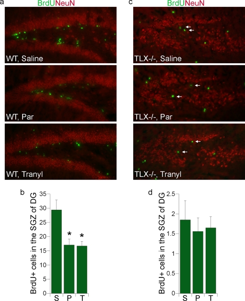
FIG. 6.
LSD1 inhibitor treatment leads to reduced cell proliferation in the dentate gyri of adult mouse brains. (a) Representative images of hippocampal dentate gyrus brain sections of wild-type adult mice that were injected with saline, pargyline (Par) or tranylcypromine (Tranyl) and treated with BrdU. Brain sections were stained with BrdU (green) to measure cell proliferation. NeuN staining (red) was included to show the structure of dentate gyrus. (b) Average numbers of BrdU-positive (BrdU+) cells in the subgranular zone (SGZ) of the dentate gyrus (DG) in one field of 20-μm wild-type brain sections. (n = 6 mice for each treatment group). S, saline; P, pargyline; T, tranylcypromine. Error bars are standard deviations of the means. *, P < 0.001 by one-way ANOVA. (c) Representative images of hippocampal dentate gyrus brain sections of TLX−/− adult mice that were injected with saline, pargyline (Par), or tranylcypromine (Tranyl) and treated with BrdU. Brain sections were stained with BrdU (green) and NeuN (red). The BrdU-positive cells that are located in the SGZ of DG are indicated by arrows. (d) Average numbers of BrdU-positive (BrdU+) cells in the SGZ of the DG in one field of 20-μm TLX−/− brain section. (n = 6 mice for each treatment group). S, saline; P, pargyline; T, tranylcypromine. Error bars are standard deviations of the means.

