
FIG. 1.
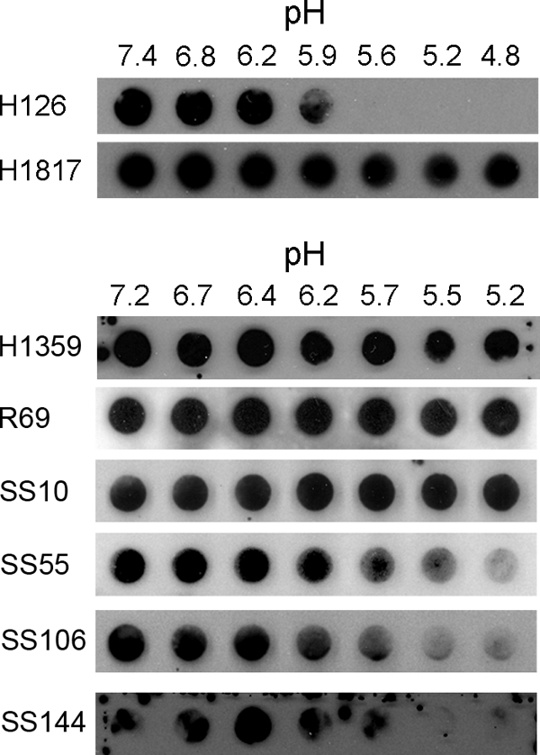
Antibody reactivity of low pH-treated virions. Extracellular HSV-1 KOS virions (105 PFU) were treated for 10 min at 37°C with medium buffered to the indicated pHs and were blotted immediately to a nitrocellulose membrane. Blots were probed at neutral pH with the indicated gB-specific antibodies, followed by horseradish peroxidase-conjugated goat secondary antibody. The exposure shown for MAb H126 highlights the pH threshold of conformational change.