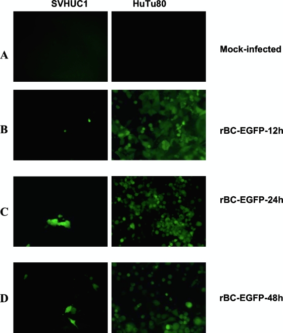
FIG. 1.
Levels of virus replication and spread of NDV differ in normal human epithelial cells and HuTu80 tumor cells. SVHUC1, normal, immortalized human uroepithelial cells, and HuTu80 intestinal epithelial tumor cells were infected with rBC-EGFP virus at an MOI of 0.01. (A to D) Left panels indicate SVHUC1 cells, and right panels indicate HuTu80 cells. (A) Mock-infected cells; (B) expression of EGFP in a few virus-infected normal cells compared with extensive EGFP fluorescence in virus-infected HuTu80 cells by 12 h postinfection; (C) absence of virus spread and replication in normal cells compared with extensive EGFP and virus spread in tumor cells by 24 h postinfection; (D) expression of EGFP in a few cells, with absence of viral spread in normal human cells at 48 h and complete destruction of a monolayer, with clumps of dead cells expressing EGFP in HuTu80 tumor cells.

