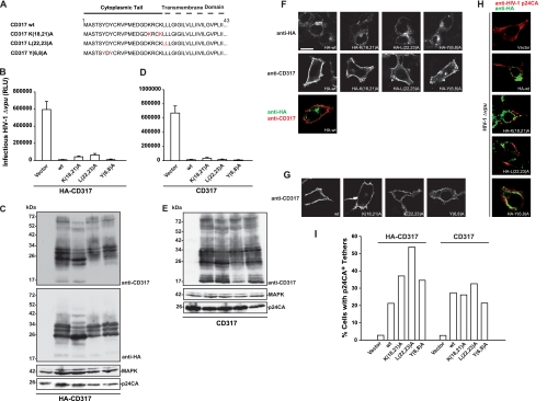
FIG. 1.
Characterization of basic properties of CD317 CT mutants. (A) Amino acid alignment of CD317 and CD317 CT mutants. (B and D) Released infectivity of 293T cells transfected with pHIV-1Δvpu and expression constructs for HA-tagged (B) or untagged (D) CD317. RLU, relative light units. Arithmetic means ± standard deviations (SD) of one experiment performed in triplicate are given. (C and E) Western blot analyses of corresponding 293T cell lysates. CD317 was detected using a polyclonal anti-CD317 antibody recognizing the extracellular domain of CD317 (16). MAPK, mitogen-activated protein kinase. (F) 293T cells grown on coverslips were transfected with expression plasmids for the indicated HA-CD317 variants. Presented are confocal micrographs for the localization of HA-CD317 proteins by using an anti-HA antibody (upper panels) or the polyclonal anti-CD317 antibody (middle panels). The bottom panel depicts a merge picture of a double staining of a 293T cell expressing HA-WT CD317 with anti-HA and anti-CD317 antibodies. Scale bar, 10 μm. (G) Micrographs of 293T cells expressing the indicated CD317 variants following staining with anti-CD317 antibody. (H) 293T cells transfected with pHIV-1Δvpu and the indicated HA-CD317 proteins were stained for HIV-1 p24CA (red) and HA-CD317 (green). (I) Quantification of p24CA/HA-CD317 double-positive cells showing virus tethering (Gag accumulation at the surface and/or Gag internalization), essentially as previously reported (4).

