Figure 5.

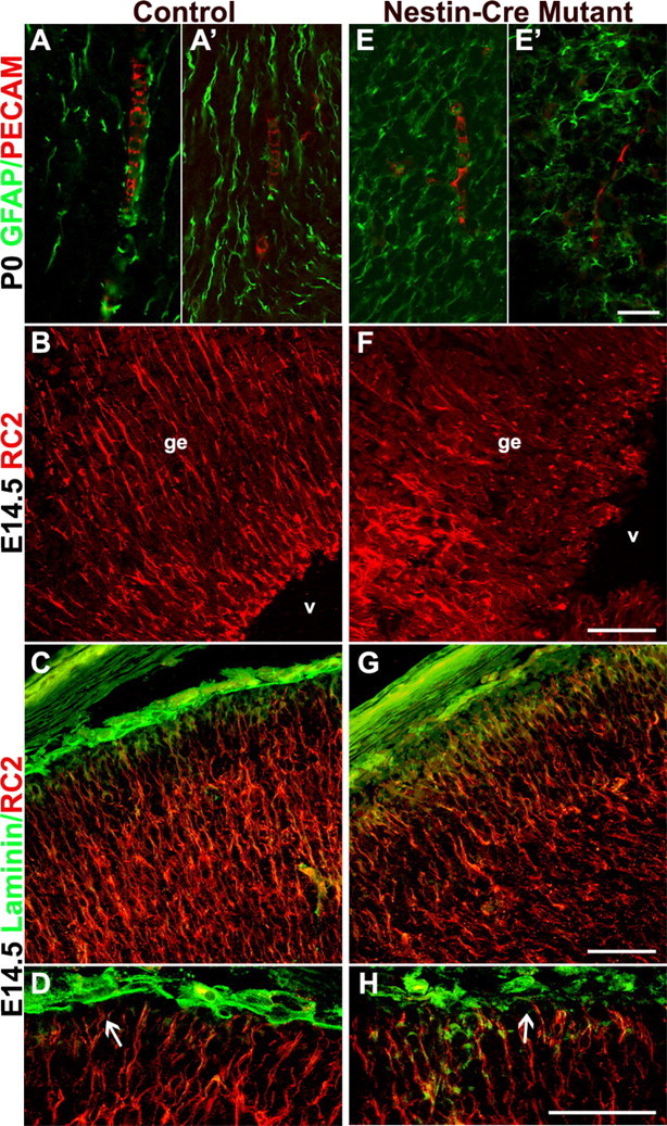
Itgβ8 nestin-cre mutants have abnormally organized cortical glia. Anti-PECAM/GFAP immunostaining of endothelial cells and glia, respectively, in P0 coronal sections of the dorsal forebrain is shown. A, A′, Control. E, E′, Nestin-cre mutant. Notice the disorganization of the astroglia and the lack of alignment between endothelial cells and the astroglial processes (E, E′). Anti-RC2 immunostaining of radial glia in E14.5 coronal sections of the ganglionic eminence is shown. B, Control. F, Nestin-cre-targeted mutant. Radial glia in F are badly disorganized, most likely because of hemorrhage within the ganglionic eminence (ge) near the lateral ventricles (v). Anti-RC2 and anti-laminin immunolabeling of E14.5 coronal sections demonstrating radial glia cell morphology in cortices of a control animal (C, D) and a nestin-cre mutant (G, H) is shown. Attachment of the radial glia to the pial surface in the nestin-cre mutant does not appear altered (H, arrow), and glial processes display normal organization. Scalebars: E′, 20 μm; F-H, 50 μm.
