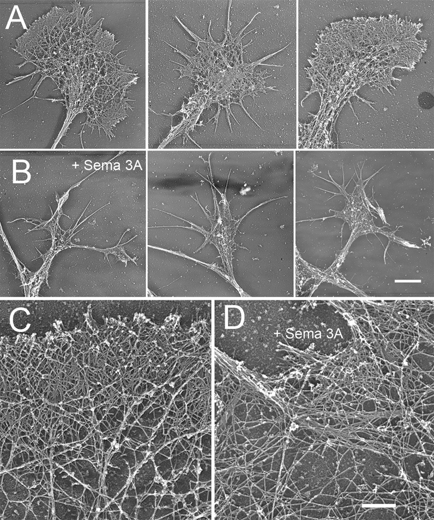
Figure 11.
Rotary shadow EM of DRG growth cones growing on high substrate bound laminin. Growth cones from both untreated (A) and Sema 3A treated (500 ng/ml, 30 min) (B) contain f-actin organized as meshworks or bundles. The amount of area containing actin meshworks appears reduced. Bar= 6 µm. (C) High magnification rotary shadow EM views showing f-actin organization of untreated growth cones on high substrate bound laminin. (D) Actin meshworks persist after Sema 3A treatment, but sometimes show larger open areas and reduced density at the leading edge. Bundled f-actin appears unchanged. Bar= 700 nm.

