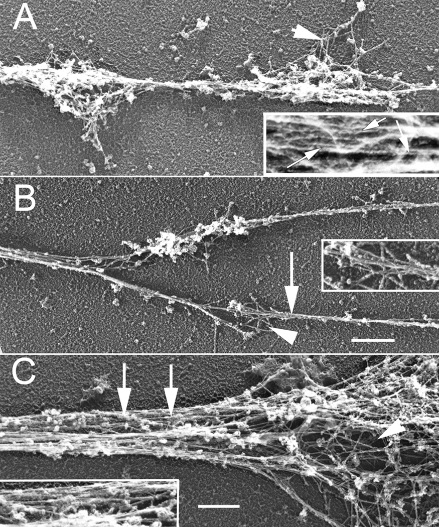
Figure 5.
Rotary shadowed EM higher magnification views of Sema 3A treated growth cones from Figure 4. (A) The neurite of a fully collapsed growth cone contains small amounts of actin meshwork (arrowhead) in varicosities and bundles (arrows) along its length (inset). (B) A partially collapsed growth cone containing two filopodia with thin f-actin bundles (arrow) and small areas of meshwork (arrowhead)(inset). (C) A partially collapse growth cone that still retains both meshworks (arrowhead) and bundles (arrows and inset). A & B, Bar= 720 nm; C, Bar= 600 nm.

