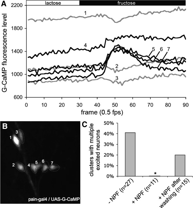
Figure 3.
NPF suppression of sugar excitation of NPFR1-positve PAIN neurons from postfeeding larvae. Stimulation paradigm: the tissue was initially perfused with HL6–lactose solution for 5 min before imaging. Each tissue was imaged in HL6–lactose for 60 s and then in HL6–fructose for 120 s. A, Representative traces showing G-CaMP fluorescence changes of thoracic PAIN neurons of normal postfeeding larvae (96 h AEL) in response to fructose. The numbers on the traces correspond to the labeled neurons in B. B, G-CaMP labeled PAIN neurons in the second thoracic segment. C, Quantification of the NPF inhibitory effect on sugar excitation of thoracic clusters of PAIN neurons. Perfusion of larval tissues with 1 μm NPF significantly attenuated the number of G-cAMP-expressing PAIN neurons excitable by fructose.

