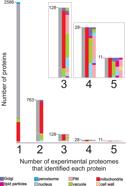
Fig. 5.
Proteins identified in multiple experimental proteomes. Each of the 3,516 identified proteins was found in one to five experimental proteomes. The bars indicate the total number of proteins found in one, two, three, four, or five proteomes. The inset shows an enlargement of the bars for the proteins identified in three, four, or five proteomes. The colors indicate in which experimental proteomics studies the proteins were identified: pink, plasma membranes; beige, cell walls; red, mitochondria; green, vacuoles; violet, Golgi apparatus; gray, nuclei; blue, peroxisomes; magenta, lipid particles.

