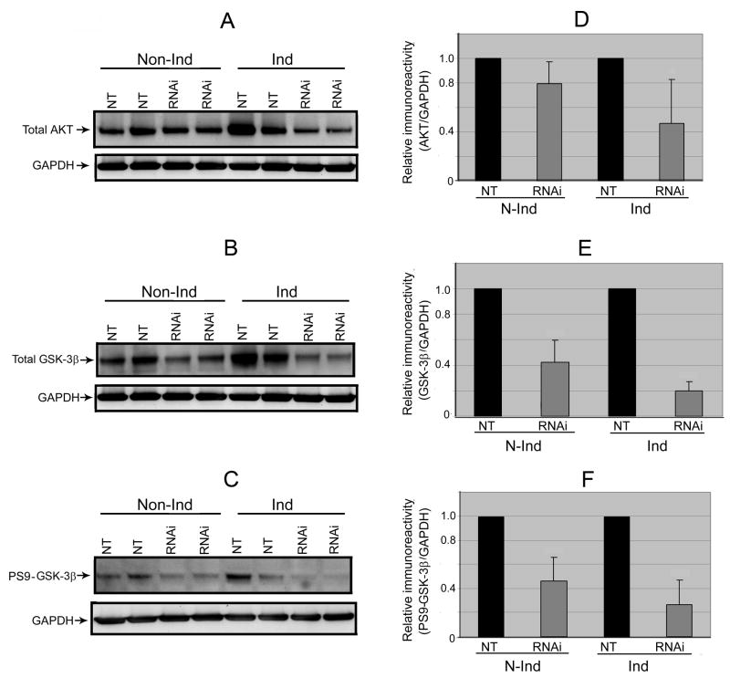
Fig. 5. Down-regulation of AKT, GSK-3β and P-GSK-3β-Ser9 from PGC-1α RNAi.
We probed lysate preparations of the differentiated 3D5 cultures, with or without TetOff induction, by immunoblotting analysis for (A) AKT and (B) GSK-3β as well as (C) its phosphorylated form (p-GSK-3β-Ser9). Immunoreactivities of AKT, GSK-3β and p- GSK-3β-Ser9appeared down-regulated following PGC-1α knockdown of non-induced cells and such reduction declined further after TetOff induction. This was verified quantitatively by densitometric analyses using GAPDH to normalize sample loading as illustrated for (D) AKT [20% reduction detectable with the non-induced (p=0.06) but 53% decrease after TetOff induction (p=0.02)], (E) GSK-3β [57% drop discernible with the non-induced (p<0.001) while 80% decline in the induced (p<0.0001)] and (F) p-GSK-3β-Ser9 [54% decrease detected in the non-induced but 74% reduction after TetOff induction (p<0.001)]. Values represent means ± SD derived from immunoblots of the PGC-1α RNAi-treated and their corresponding NT controls from 4 experiments.

