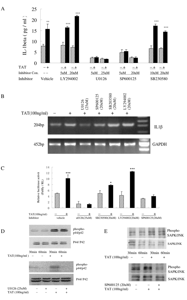
Fig 2.
Activation of p44/42 and JNK MAP kinase pathways involved in TAT-induced IL-1β release by human monocytes. A) Primary human monocytes (2 × 10⁵ cells) pretreated with U0126 (5 µM and 25 µM), SB230580 (10 µM and 20 µM), SP600125 (5 µM and 20 µM), LY294002 (5 µM and 20 µM) for 2 h were stimulat with (+) or without (−) 100 ng/ml HIV TAT for an additional 12 hours. IL-1β in cell supernatants was analyzed by ELISA. B) RT-PCR for IL-1β and GAPDH was performed on total RNA from monocytes after a 3-hour treatment with 100 ng/ml TAT following a 2-hour pretreatment with U0126 (25 µM), SB230580 (20 µM), SP600125 (20 µM) or LY294002 (20 µM). GAPDH was included as a loading control. C) THP-1 cells were transfected with the luciferase reporter constructs containing −4016 bp to +38 bp of the IL-1β promoter with an internal control Renilla luciferase reporter, RL-TK, and cultured with (+) or without (−) 100 ng/ml TAT for another 12 hours after a 2-hour inhibitor treatment. Cells were then assayed for luciferase activity. All values of the ELISA results are the means+SD of three experiments. Differences between TAT-treated cells and normal control cells were considered significant at P < 0.05. *, p<0.05; **, p<0.01; or ***, p<0.001.D and E) Cell lysate from monocytes treated with (+) or without (−) TAT (100 ng/ml) for 30 minutes and 60 minutes (up panel) or treated with TAT (100 ng/ml) for 1 hour with (+) or without (−) inhibitors U0126 (25 µM) or SP600125 (20 µM) (lower panel) were prepared and subjected to Western blotting using phospho-JNK or phospho-p44/p4 Abs. Blots were re-probed with Abs to total p42/44 or JNK as loading control. Blots shown are representative of three separate experiments.

