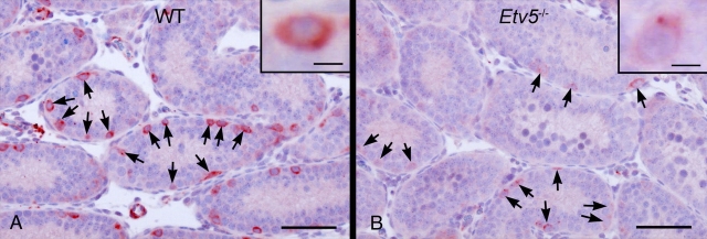
FIG. 5.
RET immunohistochemistry in WT and Etv5−/− testes (n = 4). The intensity of RET staining per spermatogonia is markedly reduced in Etv5−/− testis (B), although the numbers of cells staining for RET (arrows) are comparable to those of the WT testis (A). Insets are higher magnification of spermatogonia showing differences in staining intensity. Bar = 50 μm (10 μm in insets).

