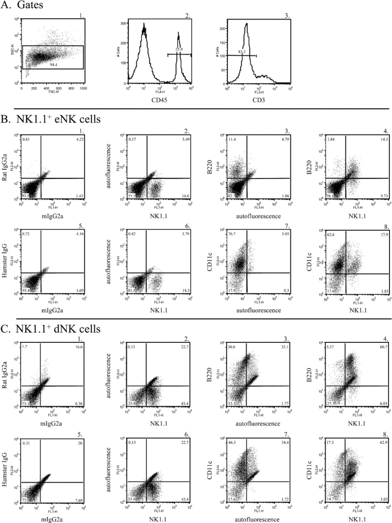
FIG. 1.

B220 and CD11c are expressed on NK1.1+ eNK and dNK cells. A) A forward-scatter (FSC) and side-scatter (SSC) gate was placed around the uterine cell population (panel A1). Next, a CD45+ gate was placed within the forward- and side-scatter gate (panel A2). A final gate was drawn within the CD45+ gate to exclude CD3+ cells (panel A3). The data depict CD45+CD3− cells. B and C) Endometrial NK cells (panel B) and dNK cells (panel C) at E10.5 were isolated and stained with either isotype control antibodies (panels B1 and B5 and panels C1 and C5), an FITC-conjugated anti-NK1.1 antibody (panels B2 and B6 and panels C2 and C6), a biotin-conjugated anti-B220 antibody (panels B3 and C3), a PE-conjugated anti-CD11c antibody (panels B7 and C7), a biotin-conjugated anti-B220 antibody and an FITC-conjugated anti-NK1.1 antibody (panels B4 and C4), or a PE-conjugated anti-CD11c antibody and an FITC-conjugated anti-NK1.1 antibody (panels B8 and C8).
