FIG. 1.
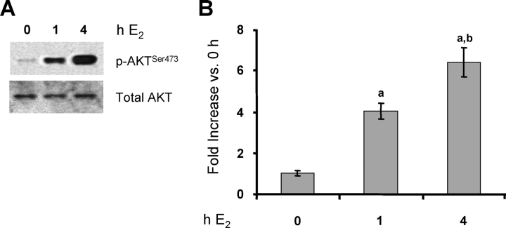
E2 activates the PI3K/AKT pathway in endometrial LE cells. Rats were treated with E2 for the indicated times or with vehicle (0 h). Total protein was extracted from LE cells, and phosphorylated and total AKT was analyzed by Western blot analysis (representative gels [A] and densitometry [B]). Densitometry results are expressed as the fold increase in phosphorylated (p)-AKTSer473 compared with control cells (0-h E2) after normalization to total AKT (mean ± SEM, n = 3 independent LE cell samples/group; each sample contained cells pooled from two uteri). a, vs. 0-h E2 (P < 0.01); b, vs. 1-h E2 (P < 0.05).

