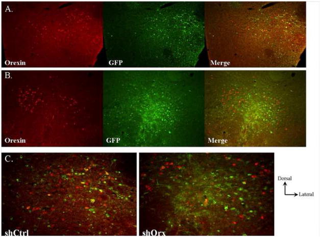
Figure 4.
Example of lateral hypothalamic infection of shCtrl (A) and shOrx (B) virus. Sections were processed for detection of orexin neuropeptide (red) and GFP from viral infection (green). Right panels in (A) and (B) show merged images. (C) Higher power analysis of infection sites showing persistent orexin expression in neurons infected with shCtrl in contrast to no orexin expression seen in neurons infected with shOrx.

