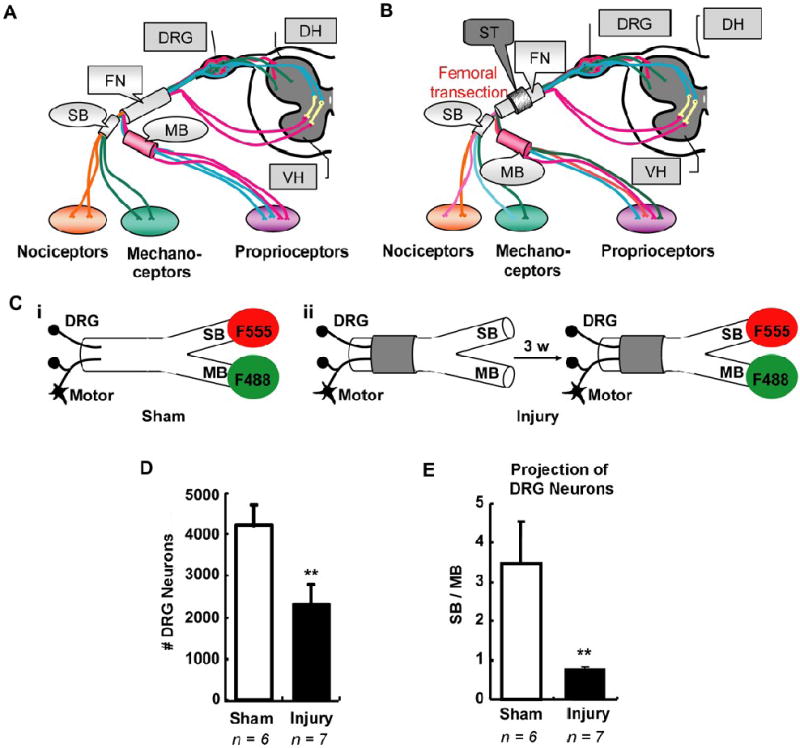
Figure 1.

Femoral transection induced abnormal sensory neuron reinnervation. A, Schematic illustration of the adult femoral nerve, which consists of parent trunk, saphenous branch (SB) and motor branch (MB). B, Schematic illustration of axon reinnervation in transected femoral nerve. Transection of parent trunk with a lesion gap in a guidance tube usually results in inappropriate axons reinnervating either SB or MB. C, Scheme of simultaneous double retrograde tracing. F555 (red fluorescent) and F488 (green fluorescent) were independently applied at either SB or MB in sham group (i) or in injury group three weeks after femoral transection (ii). D – E, Counts of labeled DRG neurons correspond to sensory neurons innervating either SB or MB. Statistical results showed the total number of retrograde labeled neurons significantly decreased after femoral transection (D). Meanwhile, their projection towards SB or MB significant changed compared to sham group, demonstrating abnormal sensory neuron reinnervation (E). DRG, dorsal root ganglion; DH, dorsal horn; VH, ventral horn; FN, femoral nerve; SB, saphenous branch; MB, motor branch; ST, silicone tubing; F555, Dextran AlexaFluor 555; F488, Dextran AlexaFluor 488; 3 w, 3 weeks. Values represent mean ± S.E.M; n = 6 – 7. ** p < 0.01 compared to sham group, analyzed by Student’s t-test.
