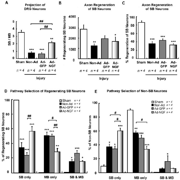
Figure 4.

Ad-NGF administration significantly increased the accuracy of SB sensory neuron reinnervation 3 weeks post-injury. Femoral transection, adenoviral administration, and sequentially triple retrograde tracing were performed as illustrated in figure 2A. L2 – L4 DRGs were processed at 3 weeks post-injury. A, The ratio of F555 to F488 labeled DRG neurons (either in the presence or absence of FG-labeling) indicate the ratio of sensory neurons with axons extending into either SB and MB. Statistical results showed Ad-NGF administration significantly increased sensory neuron reinnervation into SB, compared to non-Ad and Ad-GFP groups. B – C, The number of F555/FG+ and F488/FG+ co-labeled neurons designate the number or percent of regenerating SB neurons. Injection of adenoviruses did not significantly affect the number of regenerating SB neurons (B) as well as the percentage of regenerating SB neurons (C). D – E, Pathway selection for sensory neuron reinnervation was identified and calculated by counting of multi-labeled DRG neurons. The percentages of F555, F488, F555 & F488 labeling corresponds to the proportion of reinnervating SB, MB, and dual pathways, respectively. Injection of Ad-NGF along post-traumatic SB significantly increased the accuracy of SB neuron reinervation (D). Interestingly, non-SB neurons also preferentially grow into SB with Ad-NGF administration (E). SB, saphenous branch; MB, motor branch; non-Ad, non-adenovirus; Ad-NGF, recombinant adenovirus encoding NGF; Ad-GFP, recombinant adenovirus encoding GFP. Values represent mean ± S.E.M; n = 4. * p < 0.05; ** p < 0.01; *** p < 0.001 compared to sham group; # p < 0.05; ## p < 0.01 compared to non-Ad or Ad-GFP group, analyzed by One-Way ANOVA followed by Tukey post hoc test.
