Figure 6.

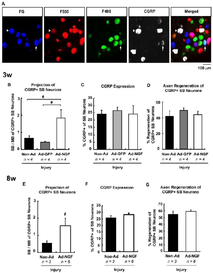
Ad-NGF administration significantly increased accuracy of CGRP-positive nociceptive neuron reinnervation. The cryosections from L2 – L4 DRG were immunostained with CGRP, a specific marker for peptidergic nociceptive axons. A, Co-labeling of FG and CGRP corresponds to CGRP+ SB neurons. F555- and F488-labeling designates neuron reinnervating SB or MB, respectively. The fluorescent images showed CGRP+ SB neurons reinnervated either SB or MB. B – D, Neuron reinnervation 3 weeks post-injury. Ad-NGF administration significantly increased the ratio of SB / MB projection of CGRP+ SB neurons (B). However, the expression of CGRP (C) and the regeneration of CGRP+ SB neurons (D) did not change significantly. E – F, Neuron reinnervation 8 weeks post-injury. The ratio of SB / MB projection of CGRP+ SB neurons significantly increased with Ad-NGF administration (E). Ad-NGF application did not influence CGRP expression (F) and regeneration of CGRP+ neurons (G). Arrowhead, CGRP+ SB neurons reinnervated SB; arrow, CGRP+ SB neurons reinnervated MB. Values represent mean ± S.E.M; n = 3 – 6. # p < 0.05 compared to non-Ad or Ad-GFP group, analyzed by Students’ t-test (between only two groups) or One-Way ANOVA followed by Tukey post hoc tests. CGRP, calcitonin gene-related peptide; SB, saphenous branch; MB, motor branch; non-Ad, non-adenovirus; Ad-NGF, recombinant adenovirus encoding NGF; Ad-GFP, recombinant adenovirus encoding GFP. Scale bar, 100 μm.
