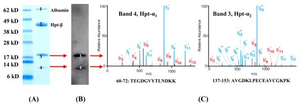
Figure 3. Antigen Identification for mAb #2.
Reduced SDS-PAGE of an affinity purified mixture (5 μg) from IgG- and albumin-depleted pooled lung cancer plasma, stained with Coomassie blue (A). Bands 1 and 2 are albumin and Hpt-β chain, respectively. Bands 3 (~18 kDa) and 4 (~10 kDa) were both recognized by mAb #2 (B) and were subsequently identified as the Hpt-α2 and Hpt-α1 chain by LC-ESI-MS (C).

