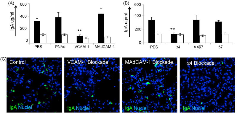
Figure 3.
Functional blockade of VCAM-1 and α4 integrins inhibits IgA ASC accumulation to the lactating mammary gland. (a) IgA levels in milk (black bars) and serum (white bars) following functional blockade of VCAM-1 (three animas), MAdCAM-1 (five animals), and PNAd (four animals) and diluent control as described in Materials and methods. (b) IgA protein levels in milk (black bars) and serum (white bars) following functional blockade of lymphocyte-expressed integrins α4 (six animals), α4β7 (five animals), β7(seven animals), and diluent control (ten animals), as listed in the X-axis. (c) Immunohistology of mammary tissue following blockade of VCAM-1, MAdCAM-1, α4 integrins and diluent control. IgA staining is shown in green, nuclei are shown in blue. Quantitative data in figures 3a and 3b represents the average value of all animals tested in each experimental group. Error bars represent the standard error of the mean. ** indicates statistical significance at p<0.01 when compared to IgA levels in animals injected with PBS.

