Fig. 4.
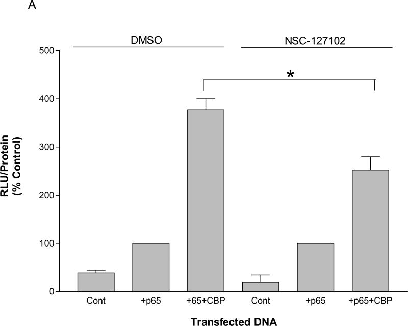
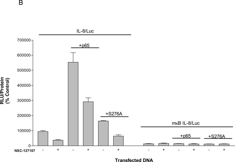
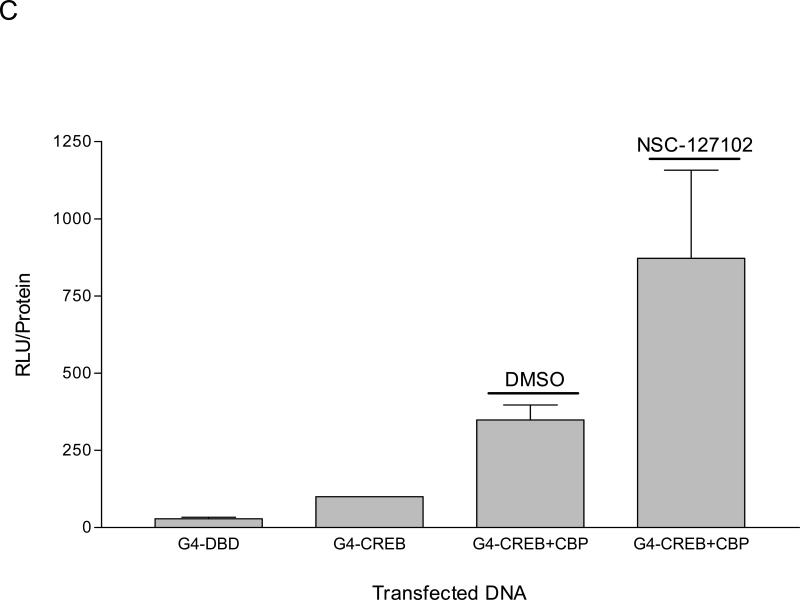
NSC-127102 inhibits activity mediated by p65 and CBP. (A) HepG2 hepatoma cells were transfected with a construct that contains five κB elements upstream of a luciferase reporter gene in the presence or absence of co-transfected p65 and CBP. Twenty-four hours later, cells were treated with 200 μM NSC-127102 for an additional 24 h. Cell lysates were prepared and luciferase activity was measured. Luciferase activity was normalized to protein concentration. The data represent the average of triplicate determinations ± S.E. (*p<0.05, Student's t test), and is representative of four independent experiments. (B) HepG2 hepatoma cells were transfected with IL-8/Luc or mκB IL-8/Luc the presence or absence of co-transfected p65 or S276A. Twenty-four hours later, cells were treated with 200 μM NSC-127102 for an additional 24 h. Cell lysates were prepared and luciferase activity was measured. Luciferase activity was normalized to protein concentration. The data are representative on three independent experiments. (C) HepG2 cells were co-transfected with a construct that has CREB fused to the Gal4 DNA binding domain (G4-CREB) and a construct that has five Gal 4 DNA binding sites upstream of the E1B TATA box and luciferase reporter gene ((GAL4)5E1bLuc). After 24 h, cells were treated with 200 μM NSC-127102 for an additional 24 h. Cells were subsequently harvested, as described in panel A, and luciferase activity was measured and normalized to protein concentration.


