
Fig. 1.
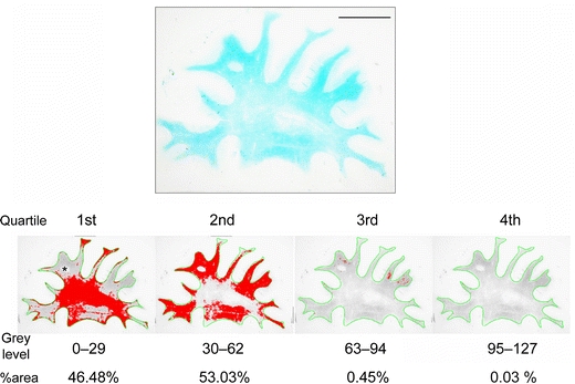
Quartile analysis of LFB-stained section from the frontal lobe of a VaD patient (74 years). The WM was outlined in green and the grey levels within the WM were divided into four quartiles. The area percentage (%area) for each quartile was calculated (labelled in red). Myelin index was then calculated for each case and control (see “Methods”) (scale bar 1 cm)