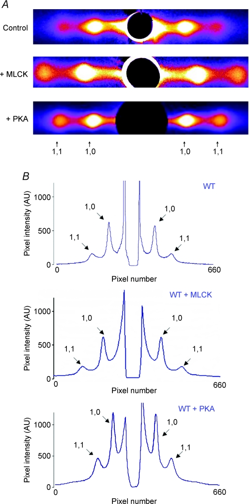
Figure 3.
Effects of MLCK and PKA treatments on intensities and spacings of 1,0 and 1,1 equatorial peaks in WT skinned myocardium A, X-ray diffraction patterns from skinned myocardium that was untreated (control; top panel) or treated with either MLCK (middle panel) or PKA (bottom panel). The ratio of intensities of the 1,0 and 1,1 equatorial reflections can be used to estimate shifts of cross-bridge mass from the region of the thick filament to the region of the thin filament. B, representative intensity traces along the equator of X-ray patterns of skinned myocardium that was untreated (control; top panel) or treated with either MLCK (middle panel) or PKA (bottom panel). The pixel intensity along the y-axis is labelled from 0 to 1000 arbitrary units and the peak profiles were not otherwise modified.

