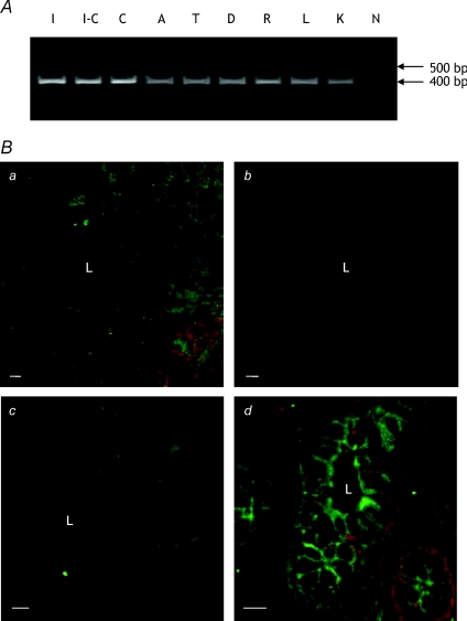
Figure 4. Expression and localisation of GLYT1 in human large intestine.
A, agarose gel elctrophoresis of products from PCR amplification of human intestinal cDNAs using GLYT1-specific primers. A single PCR product of ∼400 bp is visible for each sample. Lanes are: I, ileum; I-C, ileo-caecum; C, caecum; A, ascending colon; T, transverse colon; D, descending colon; R, rectum; L, liver; K, kidney; N, negative control (ultra-pure water replaced cDNA in the reaction). B, CLSM images of frozen sections of human descending colon stained for GLYT1. a, optical section (low power view) showing GLYT1 expression in both apical and basal membranes of cells throughout the colonic crypt. c–d, optical sections (high power views), again showing GLYT1 expression in both apical and basolateral membranes of cells in the crypt base. In the absence of anti-GLYT1 antibody (b) staining by the AlexaFluor 488-conjugated detecting antibody is negligible. a and b, which show the same region of consecutive sections, were collected and displayed using identical parameters. L, crypt lumen. Bar = 50 μm (a and b); 10 μm (c and d).

