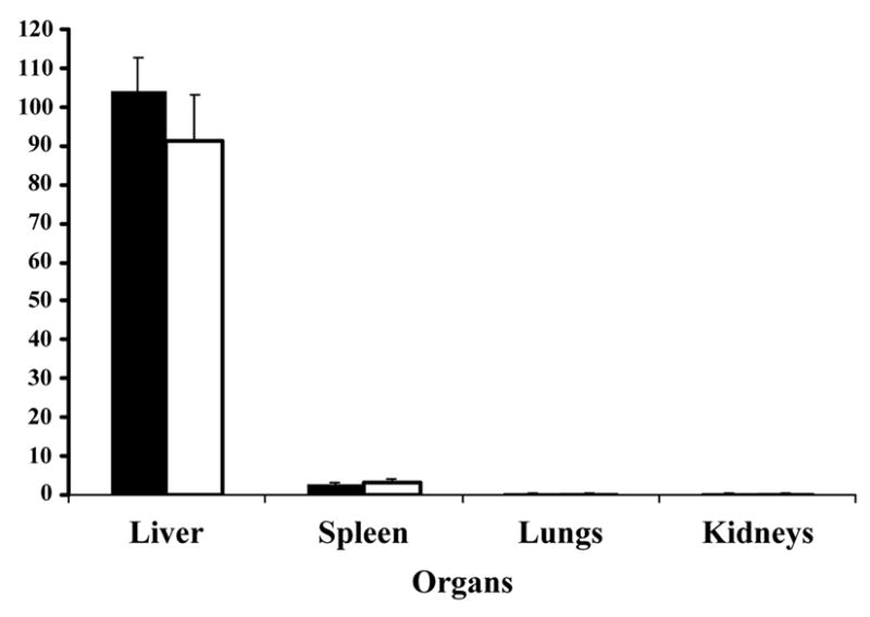
Figure 3.

Bio-distribution of CPMV in vivo in mice. CPMV-particles decorated with Gd(DOTA) on the exterior (solid bars) or Gd+3/Tb+3 ions on the interior (white bars) of the virus capsid were injected intravenously into mice. Various organs as indicated were collected after at least 30 minutes and analyzed for metal content to quantitate the amount of virus.
