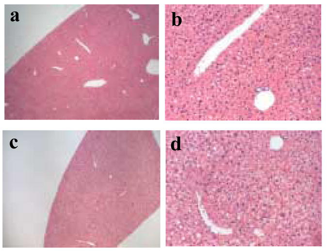
Figure 4.

Histological examination of liver tissue. Hematoxylin and eosin stained sections of liver from a saline injected mouse (panel a at 4x and panel b at 20x magnification) and a mouse injected with CPMV particles (panel c at 4x and panel d at 20x magnification).
