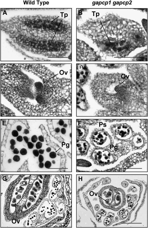
Figure 4.
The tapetal cell layer is disorganized in gapcp1gapcp2. The micrographs show transverse sections of anthers and carpels at early (top) and late (bottom) developmental stages (stages 5 and 6 and stages 8–10). A and E, Wild-type anthers; B and F, gapcp1gapcp2 anthers; C and G, wild-type carpels; D and H, gapcp1gapcp2 carpels. Ov, Ovule; Pg, pollen grain; Ps, pollen sac; Tp, tapetum. Bars = 50 μm for A to D and F and 250 μm for G and H.

