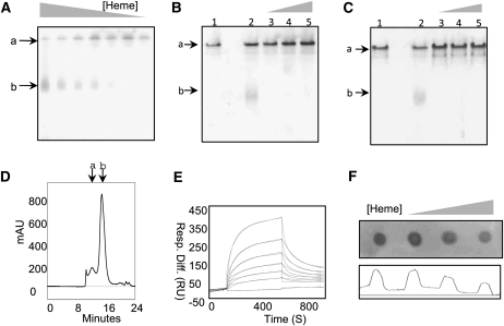
Figure 4.
Analysis of hemin binding with LS-24. A to C, Mobility shift assay of LS-24 in the presence of decreasing concentrations of hemin (A), increasing concentrations of protoporphyrin IX (B), and increasing concentrations of hematoporphyrin (C). Bands a and b correspond to LS-24 dimer and monomer, respectively. Lanes 1 and 2 correspond to native LS-24 and LS-24 preincubated with hemin (B and C). D, Size-exclusion chromatogram of heme-bound LS-24, elucidating the monomerization of LS-24 on heme binding. Peaks a and b correspond to dimeric and monomeric forms of LS-24, respectively. mAU, Milli absorbance units. E, Binding analysis by SPR of LS-24 interaction with hemin. Resp. Diff., Response difference; RU, response units. F, Dot-blot analysis presenting a decrease in anti-spermine antibody binding when LS-24 is preincubated with increasing concentration of hemin.

