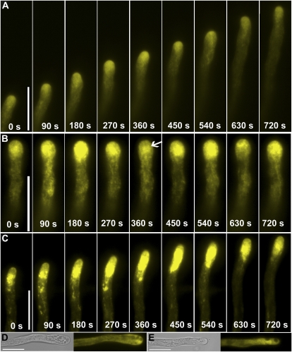
Figure 3.
Time-lapse images of transgenic pollen tubes expressing YFP-RabA4b. A, Control. B, A pollen tube treated with BFA. Arrow indicates the RabA4b labeling below apex after BFA treatment, which resembles so-called BIA. Serial images were taken starting after 20 min incubation with BFA. Therefore time point 0 s is after 20 min treatment with BFA. C, A pollen tube treated with wortmannin. Serial images were taken starting after 60 min incubation with wortmannin. Therefore time point 0 s is after 60 min treatment with wortmannin. D, A representative pollen tube treated with BFA for 60 min. E, A representative pollen tube treated with wortmannin for 90 min. For D and E, left section, transmitted light; right section, YFP channel. Scale bars = 20 μm. [See online article for color version of this figure.]

