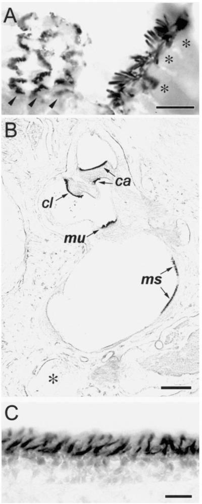
Figure 1. Affinity Purified Espin Antibody Labels Stereocilia on Cochlear and Vestibular Hair Cells in Cryosections of Decalcified Specimens from Adult Mouse and Rat (Immunoperoxidase Method).
(A) Mouse cochlea. The collections of stereocilia on the single row of inner hair cells (asterisks over hair cell bodies) are sectioned roughly longitudinally, whereas those on the three rows of outer hair cells (arrowheads) are sectioned tranversely at different levels near their base. Bar, 10 μm.
(B) Rat vestibular system, low magnification view. The collections of stereocilia on the patches of hair cells that constitute the macula utriculi (mu), macula sacculi (ms) and the cristae ampullares of the anterior (ca) and lateral (cl) semicircular canals are shown. (Asterisk, posterior semicircular canal). Bar, 300 μm.
(C) Rat vestibular system, higher magnification view of the macula utriculi. Bar, 20 μm.

