
Figure 2. Affinity Purified Espin Antibody Labels Hair Cell Stereocilia in Whole Mounts of Isolated Cochlear Sensory Epithelium from a 13-day Chicken Embryo (Confocal Immunofluorescence Method).
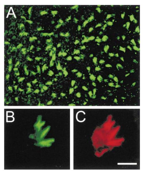
(A) Low magnification view from above the epithelium. Bar in (C), 28 μm.
(B) Higher magnification view showing several clumps of labeled stereocilia emanating from the apex of a single hair cell. Bar in (C), 5 μm.
(C) Double labeling of the same hair cell shown in (B) with rhodamine-phalloidin reveals the intensely labeled bundles of F-actin at the cores of the clumped stereocilia emanating from a less intensely labeled cuticular plate. Bar, 5 μm.