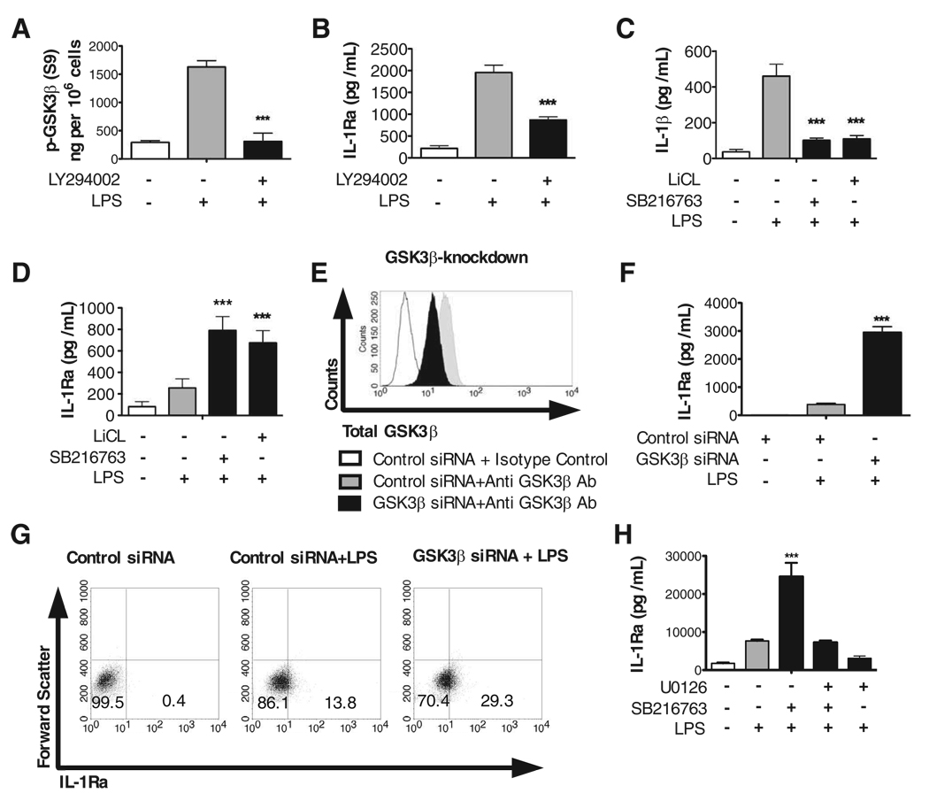
FIGURE 1.
Inactivation of GSK3 augments IL-1Ra levels, and this effect is attenuated upon ERK inhibition. A, Phospho-GSK3-β levels were determined from whole-cell lysates obtained from monocytes (5 × 10⁶ cells) that were pretreated for 2 h with the PI3K inhibitor LY294002 (25 µM) and then stimulated for 30 min with or without LPS (1 µg/ml). B, Monocytes were pretreated with the PI3K inhibitor LY294002 (25 µM) for 2 h and then stimulated with LPS. C and D, Human monocytes were pretreated for 2 h with the GSK3 inhibitor SB216763 (10 µM) or LiCl (10 mM) and then stimulated with LPS. E, siRNA-mediated knockdown in GSK3β levels was determined by flow cytometry 96-h post transfection. F and G, Human monocytes were transfected with GSK3β-specific or control siRNA for 72 h followed by the addition of LPS (1 µg/ml). H, Human monocytes were pretreated for 2 h with the GSK3 inhibitor SB216763 (10 µM) and/or the MEK1/2 inhibitor U0126 (50 µM) followed by stimulation with LPS (1 µg/ml). Unstimulated cells were treated with NaCl (10 mM) or DMSO (0.1%) as the osmolality or organic solvent controls for LiCl or SB216763, respectively. Cell-free supernatants were collected 20-h post LPS-stimulation and assayed for IL-1Ra or IL-1β levels by ELISA. ***, Statistically significant differences at p < 0.001, as compared with LPS-stimulated cells. Data represent the mean ± SD of three separate experiments.

