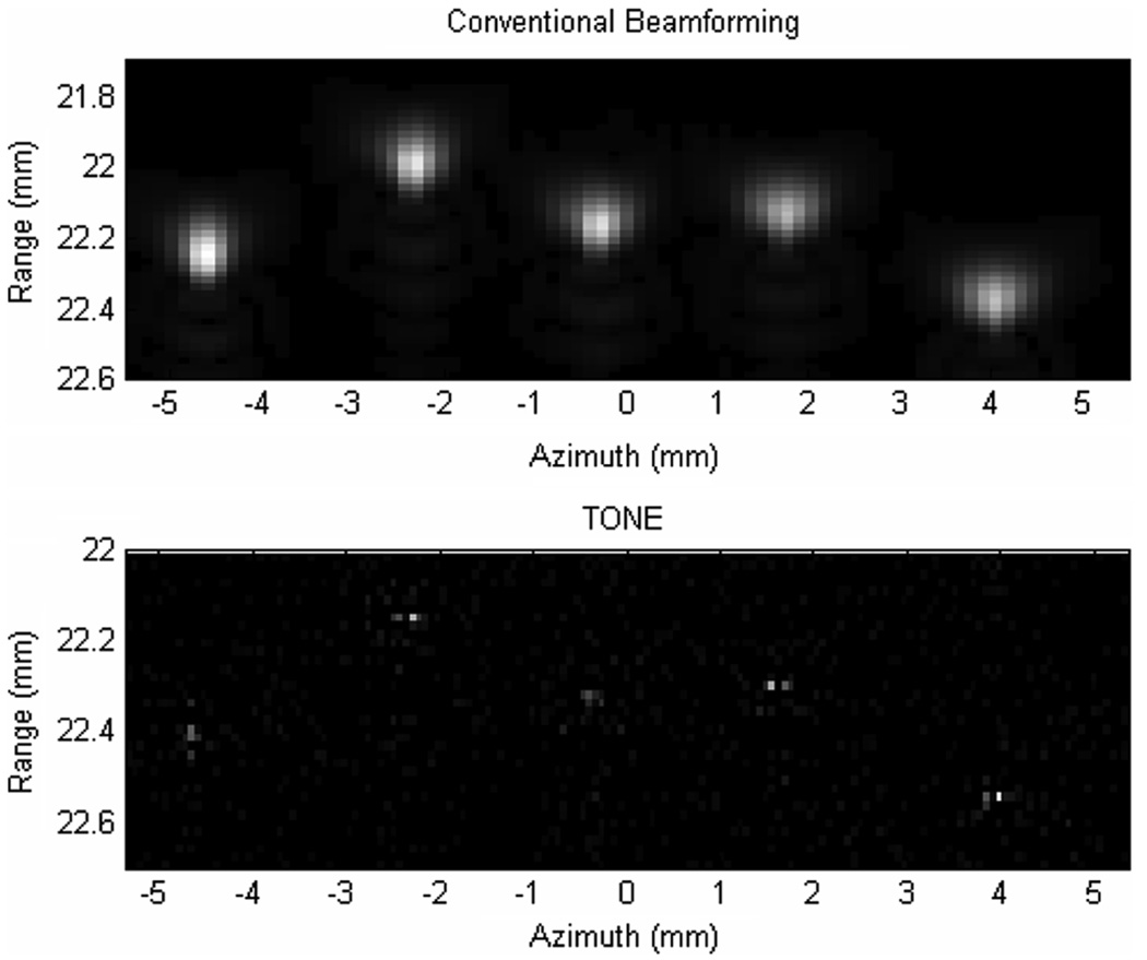
Fig. 7.
Experimental comparison of conventional beamforming (top) and TONE beamformed (bottom) images of a set of five 20-µm-diameters stainless steel wires suspended in a water tank. In the case of TONE hypothetical targets were placed every 19 µm axially and every 67 µm laterally. Images are displayed on a linear brightness scale.

